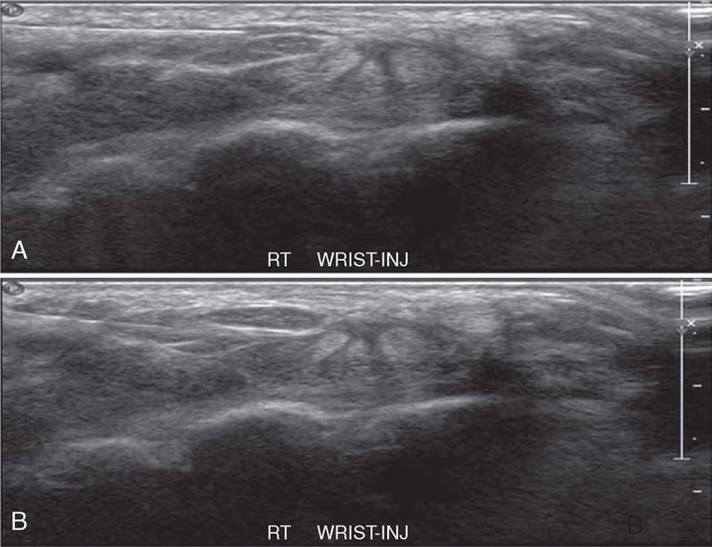
FIGURE 2.

Transverse sonogram of the right carpal tunnel in a patient with idiopathic CTS. A 27-gauge needle is shown passing from the ulnar aspect of the carpal tunnel to a position adjacent to the median nerve. (A) After positioning the needle tip next to the nerve, the local anesthetic-corticosteroid mixture is injected in order to peel the nerve off the overlying flexor retinaculum via hydrodissection. (B) Anechoic injectate is shown surrounding the deep surface of the median nerve and separating it via hydrodissection from the more deeply positioned hyperechoic flexor tendons and associated synovium. CTS = carpal tunnel syndrome.
