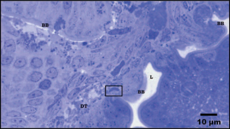
FIGURE 1.

Semi-thin section from duodenal biopsy shows panoramic view of different cutting planes. Several areas of the epithelium with normal appearance and intact trophozoite within the damaged submucosa are shown. BB: Brush border; DT: Damage tissue. Stain toluidine blue.
