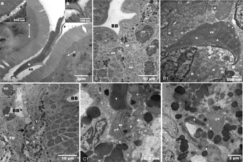
FIGURE 2.

Transmission electron micrographs are showing intraepithelial trophozoites from a patient biopsy. A: Giardia trophozoite attached on the normal duodenal brush border, (N) nucleus, (AD) adhesive disc and (F) flagellum. There is a remarkable integrity of the intestinal intercellular tight junctions (arrows). Inset (a) shows small vacuoles between cellular microvillus and the plasma membrane of the trophozoite (arrows), suggesting a biochemical interaction. Inset (b) shows that the microvilli and ventral disc interactions give rise to an electron-dense zone. B: Panoramic of the duodenal epithelium with a brush border (BB) of normal appearance. At the level of the enterocyte nuclei (EN) and goblet cell (GC) there is a trophozoite (T). Note the electron-dense granules dispersed in the tissue (arrows). B1: Higher magnification shows the normal architecture of the trophozoite, which appears to be attached with its adhesive disc (AD) to one nucleus. Note the vacuoles (V) in the normal plasma membrane, endoplasmic reticulum (ER), axonemes (A), nucleus (N), lateral crest (LC), and ventrolateral flange (VLF). Lysed cells were found near the adhesive disc. Several damaged mitochondria (M). C: Low magnification of the duodenal epithelium. There is damage in the epithelial tissue and in the brush border (BB∗), which is the likely site of entry of the trophozoites (T). We clearly see 4 trophozoites surrounded by electron-dense granules (arrow). In another area, the brush border appears intact (BB). C1: High magnification shows an intact trophozoite (T) and another trophozoite with total lysis of the dorsal membrane (T∗), and fragmentation of it adhesive disc (AD). Nearby, there are eosinophil sombrero vesicles (arrows), granules (arrowheads), and Golgi bodies (G). C2: Two Giardia trophozoites surrounded by granules (arrowheads); one looks swollen (T∗), and the other has a normal morphology (T).
