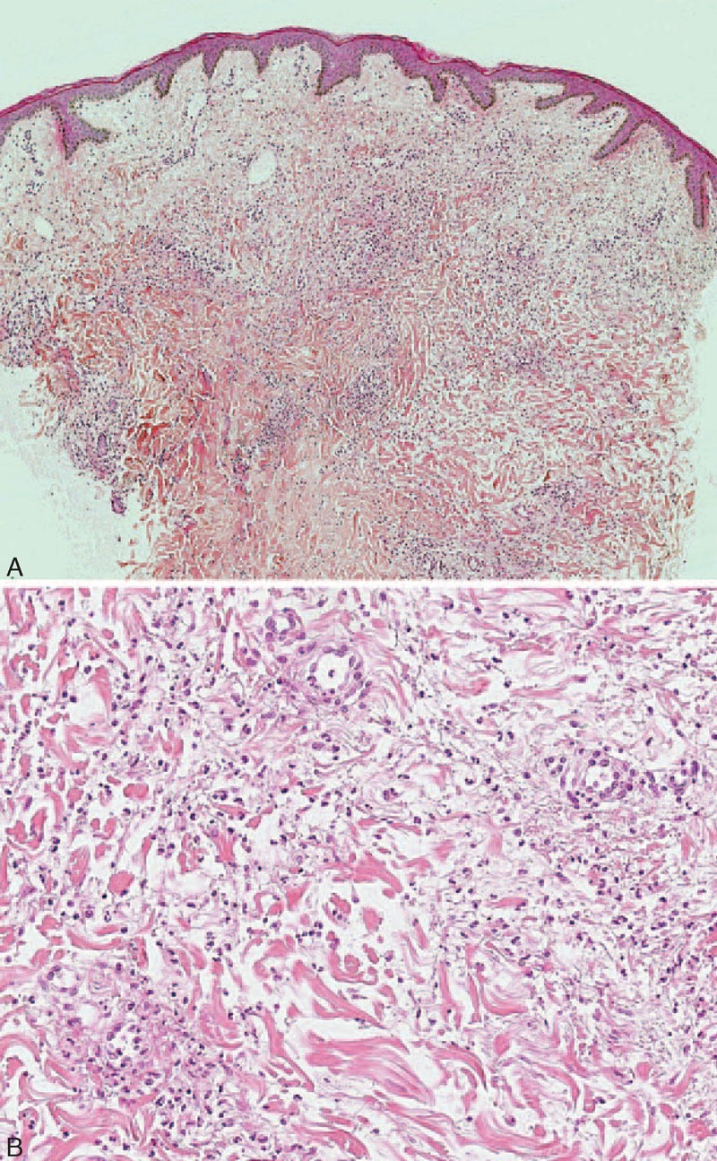
FIGURE 6.

(A) Interstitial diffuse infiltrate of neutrophils associated with leukocytoclastic debris, without vasculitis suggestive of neutrophilic urticarial dermatosis (case 3) (hematoxylin–eosin stain; original magnifications, 40). (B) Intradermal diffuse neutrophilic infiltrate (case 3) (hematoxylin–eosin stain; original magnifications, 200).
