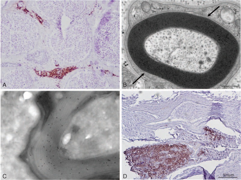
FIGURE 1.

(A) Frozen transverse section of the sural nerve of patient 1 stained with anti-CD20 antibody. Several vessels in the epineurium are surrounded by B-cell infiltrates. (B) Electron micrograph of tranverse section of the sural nerve of patient 1 showing the typical widening of the most external myelin lamellae. (C) Immunoelectron micrograph of patient 2. Anti-kappa light chain immunogold staining shows that the monoclonal protein of the patient has infiltrated the myelin sheath. (D) Frozen transverse section of the sural nerve of patient 3 stained with anti-CD20 antibody. There is a massive infiltrate of B-cells in the epineurium.
