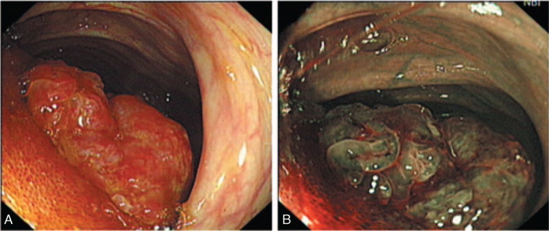
FIGURE 2.

Endoscopy appearance of GI PEComa (case 2). White light (A) and narrow band imaging (B) endoscopy showed a 2-cm diameter, polypoid tumor protruding into the lumen of the terminal ileum. GI PEComa = perivascular epithelioid cell tumors of gastrointestinal tract.
