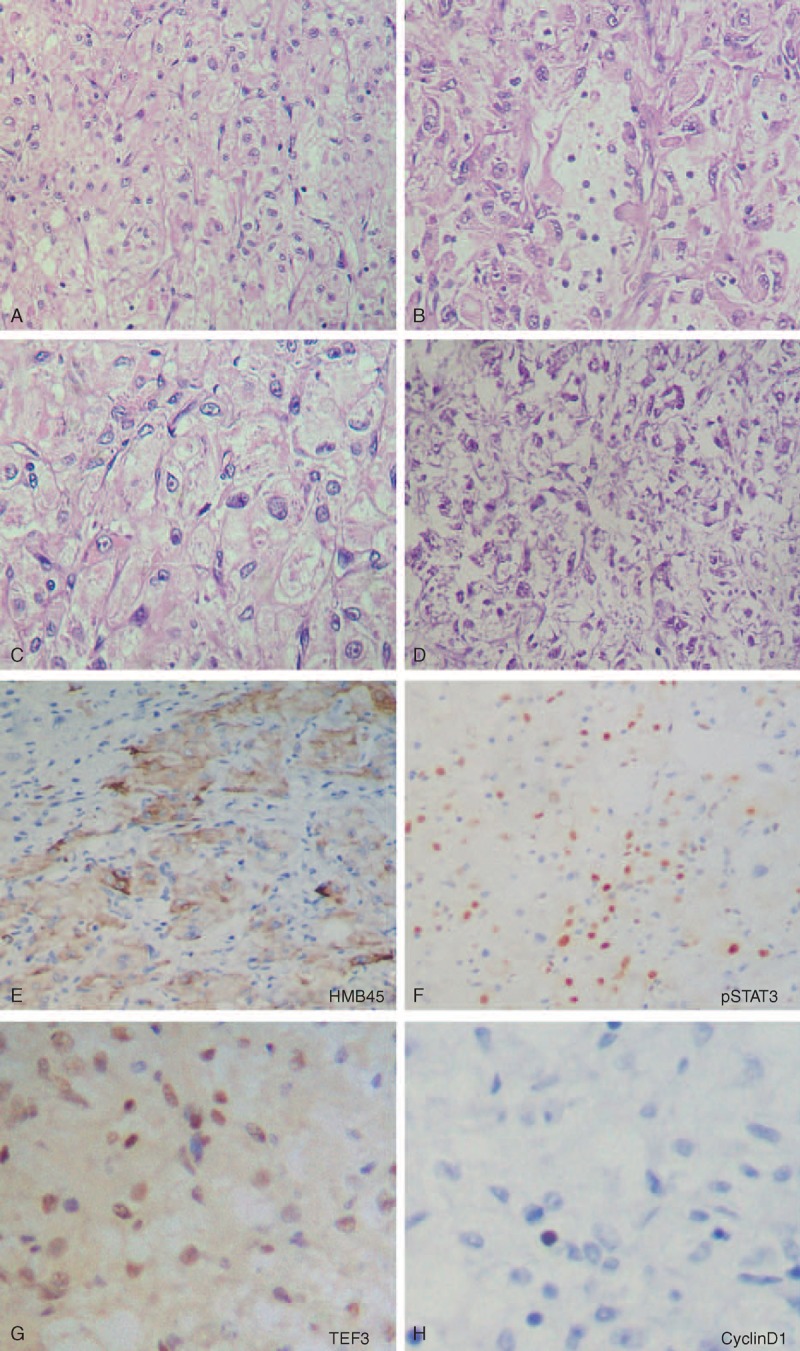
FIGURE 4.

Microscopic features of the ileum tumor (case 1). The tumor consisted of an epithelioid cell proliferation with a vaguely nested pattern (A). In some areas, the tumor displayed a pseudoglandular histological appearance (B). The perivascular epithelioid cells had clear to eosinophilic granular cytoplasm with some slightly irregular nucleus nuclei (C). Foci of coagulation necrosis were also found in the tumor (D). The tumor cells were positive for HMB45 (E), pSTAT3 (F), and TFE3 (G), and negative for Cyclin D1 (H).
