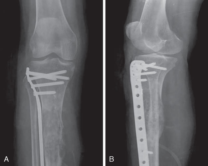
FIGURE 2.

A 50-year-old female with malignancies in FD on the right proximal tibia with a history of operation (case 1). Anteroposterior plain film (A) and lateral plain film (B) show ill-defined osteolytic lesion with cortical destruction, periosteal reaction, and ossification. No “ground-glass” opacity can be found. FD = fibrous dysplasia.
