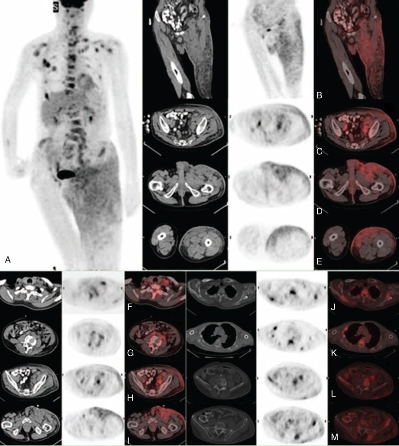
FIGURE 2.

Whole-body FDT PET/CT images demonstrated the presence of multiple FDG-avid lesions in the (A) anteroposterior 3D-MIP. The selected (B) coronal and (C–M) transaxial images revealed mild FDG uptake at the extensive cutaneous lesion with (B–E) subcutaneous invasion, involvement of (F–I) regional lymph nodes, and (J–M) multiple intense FDG-avid of skeletal metastases. 3D-MIP = 3-dimensional maximum intensity projection, FDG = fluorodeoxyglucose, PET/CT = positron emission tomography/computed tomography.
