
FIGURE 1.
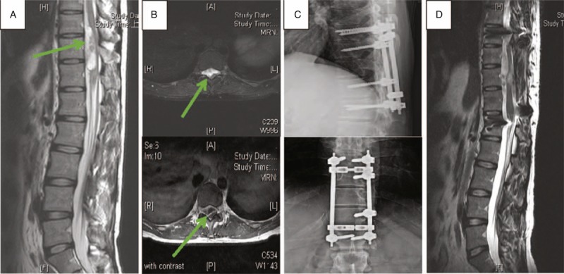
Radiological studies of Case 1. The green arrows mark the position of tumor. (A) Preoperative sagittal T2-weighted imaging of T-spine magnetic resonance imaging (MRI). (B) Preoperative axial imaging of T-spine MRI. Upper image is T2 weighted and lower image is Gd-enhanced T1 weighted. (C) Postoperative T-spine X-ray anteroposterior and lateral view. (D) Sagittal T2-weighted imaging of T-spine MRI at 2 y postsurgery.