
FIGURE 1.
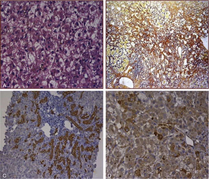
Liver histology. (A) Hepatocytes obviously displayed ballooning degeneration, necrosis with a ground glass aspect. Cholestasis and cholangiectasis also can be observed (HE 1×400). (B) There was marked periportal and septal fibrosis with moderate pericellular fibrosis (Silver staining). (C) CK19 was highly expressed in portal areas and hepatic parenchyma (1×200). (D) Immunohistochemical staining for HCV-Ag showed significantly positive.