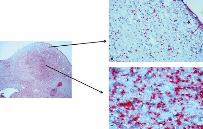
FIGURE 4.

Serotonin-immunopositive fibers in the IC showing the typical dot-like varicosities. They are less diffuse in the cortex (A) than in the central nucleus (B). Note the general immunonegativity of the neuronal bodies. In (C) Microphotograph of the inferior colliculus indicating where the images in (A) and (B) came from. Control infant (6 months old). 5-HT immunohistochemistry. Magnification (A) and (B) 40× and (C) 4×.
