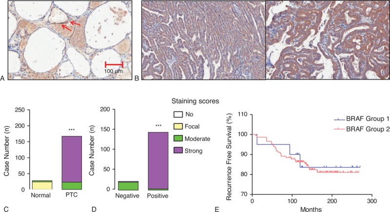
FIGURE 3.

Relation of B-RAF expression with B-RAFV600E mutation and recurrence-free survival. (A) Representative image of IHC-P using anti-B-RAF antibody in normal thyroid tissue (original magnification ×200). Arrows indicated focal staining intensity of B-RAF. (B) Representative images of B-RAF IHC-P in PTC (original magnification ×200). (C) Comparison of B-RAF expression between normal thyroid tissues and PTC. (D) B-RAF expression status according to the absence or presence of B-RAFV600E mutation. IHC staining was scored as described in the Methods (n = 167). Group comparisons were performed by linear-by-linear association. (E) Kaplan–Meier estimates of recurrence-free survival according to BRAF expression. Group 1 indicates patients with PTC showing moderate staining of BRAF; Group 2, indicates strong staining intensity. IHC = immunohistochemical staining, PTC = papillary thyroid cancer.
