FIGURE 1.

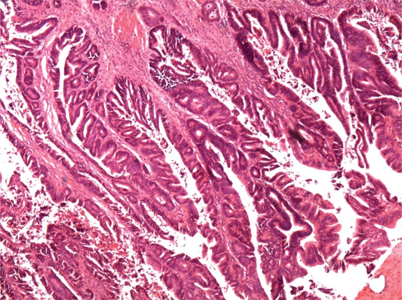
Histological appearance of a gallbladder papillary adenocarcinoma. Microscopically, these tumors present with papillary proliferation of epithelial cells and delicate fibrovascular stalks (hematoxylin and eosin staining × 40).

Histological appearance of a gallbladder papillary adenocarcinoma. Microscopically, these tumors present with papillary proliferation of epithelial cells and delicate fibrovascular stalks (hematoxylin and eosin staining × 40).