
FIGURE 1.
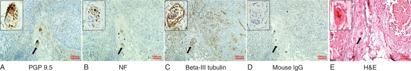
Nerve fibers present in breast cancer. (A) PGP9.5. (B) NF. (C) Class III-β-tubulin. (D) Isotype-matched antibody, mouse IgG. (E) H&E staining. Represented images of nerve fibers in breast cancer specimens. Nerve fibers were detected in serial sections of breast cancer tissues using IHC staining with 3 different specific neuronal markers. Original magnifications: 100× for the wild view; 400× for the left up corner. Scale bar, 100 μm. H&E = hematoxylin–eosin, IgG = immunoglobulin G, IHC = immunohistochemical, NF = neurofilament, PGP9.5 = protein gene product 9.5.