
FIGURE 4.
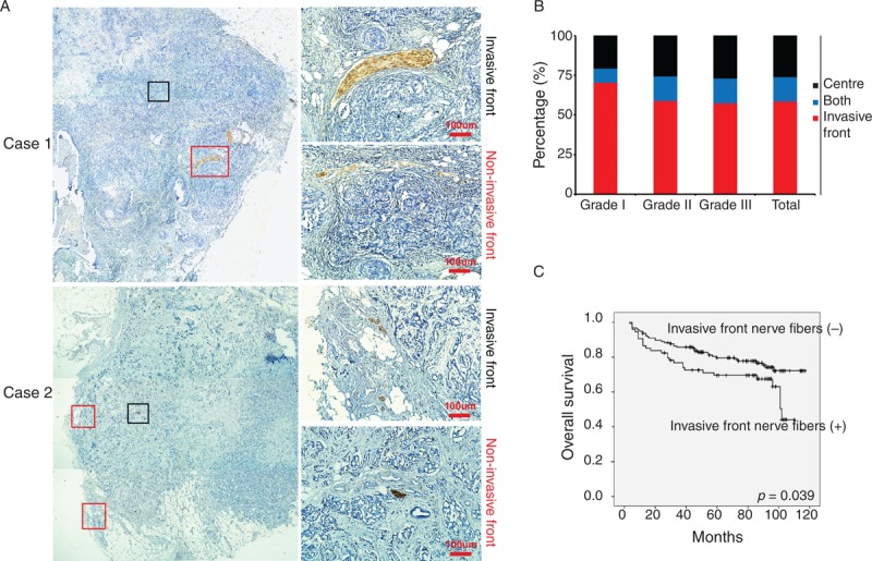
Nerve fibers in breast cancer specimens have different location. (A) Represented images of nerve fibers located in invasive front of breast cancer and the center of breast cancer. Original magnifications: left panel: 40×; right panel: 400×. Scale bar, 100 μm. (B) The proportion of nerve fibers located in invasive front and the center of tissue specimens from different grades of breast cancer. (C) Kaplan–Meier survival curve for patients with nerve fibers located in invasive front and the center of breast cancer.