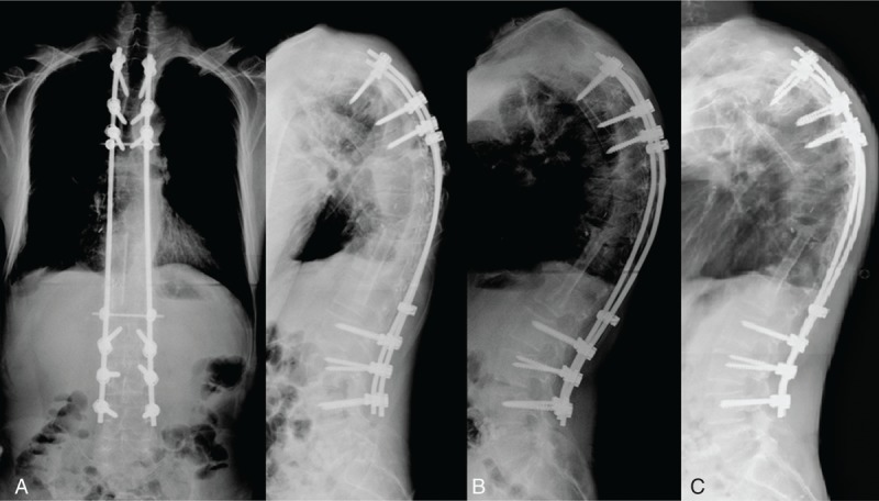
FIGURE 6.

(A) Radiograph showing better alignment after single-stage combined anterior and posterior surgeries for complicated infectious spondylitis. (B) Bony incorporation between the implanted fibular allograft and host vertebral body was noted on the lateral radiograph 1 year later. (C) The radiograph 3 years later revealed good anterior interbody fusion and acceptable sagittal alignment without hardware breakage.
