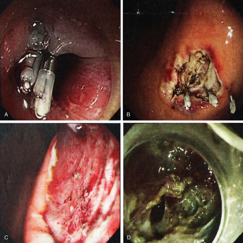
FIGURE 2.

(A and B) Images of wound surfaces were closed with titanium clips after excision of lesion. (C) Endoscopic view of wound surface tumor; no tumor tissue was left under the endoscopic inspection. (D) Gastric perforation occurred during the operation because of obvious adhesion to the serosal layer of the stomach.
