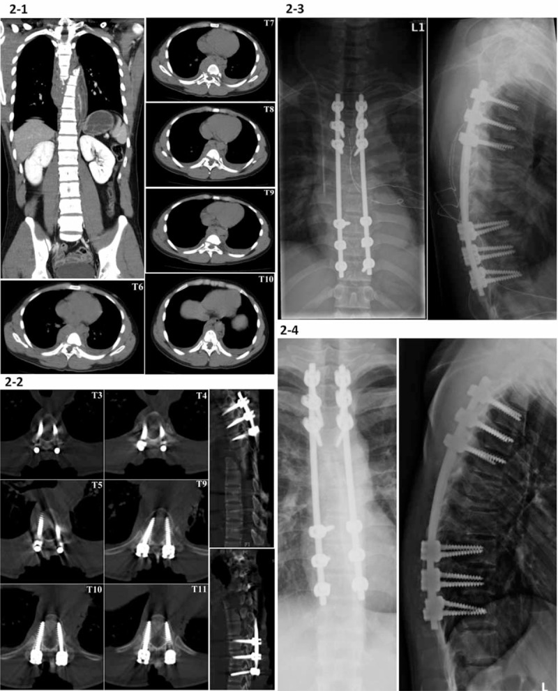
FIGURE 2.

A 23-year-old male patient sustained a complete fracture-dislocation of T6 to T10 (AO type C1) with spontaneous neurological decompression. (2-1) The preoperative chest and abdominal CT images were obtained at a regional hospital. We performed posterior instrumentation of T3 to T9 using TPS fixation under iCT navigation, except for the excessively displaced T6 to T8 vertebral bodies. (2-2) A confirmation CT scan shows good positioning of all pedicle screws. The posterior laminae from T3 to T9 were decorticated and fused with autogenous iliac cancellous bone graft and bone graft substitutes. (2-3) The postoperative radiographs show posterior instrumentation of T3 to T9 for the complete fracture-dislocation of T6 to T10. (2-4) Radiographs demonstrate good stability of the affected thoracic spine without kyphotic change or implant failure at 1-year follow-up. CT = computed tomography, iCT = intraoperative computed tomography, TPS = transpedicular screw.
