
FIGURE 3.
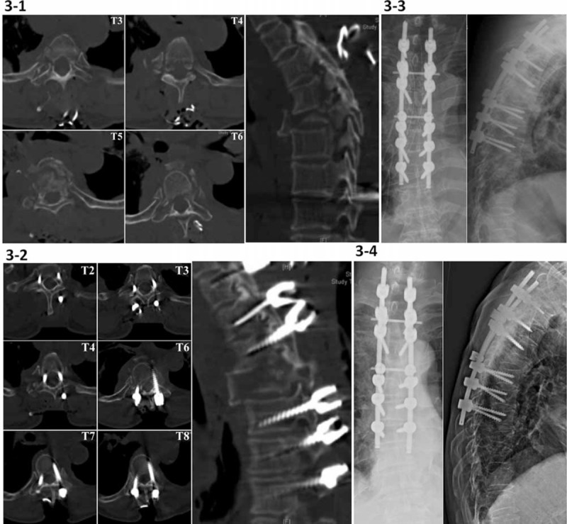
(3-1) Intraoperative spinal CT scan shows a fracture-distraction and torsion injury of T5 and multiple rib fractures and bilateral pedicle and lamina fractures involving T3 to T6 (AO type C2) in a 70-year-old female patient with ASIA grade C. TPS fixation 3 levels above and below the T5 level is guided under the iCT navigation. (3-2) The confirmation CT scan shows a grade I lateral cortical breach of right T3 TPS (1.3 mm), which did not require revision due to an acceptable screw position. After a decompression laminectomy at the T5 level, autogenous bone chips and bone substitutes were placed on the decorticated laminae from T2 to T8 for posterior fusion. (3-3) The postoperative radiographs show posterior instrumentation of T2 to T8 for the complete fracture-distraction of T3 to T6. (3-4) There is no kyphotic collapse at the 1-year follow-up radiographic examination. ASIA = American Spinal Injury Association, CT = computed tomography, iCT = intraoperative computed tomography, TPS = transpedicular screw.