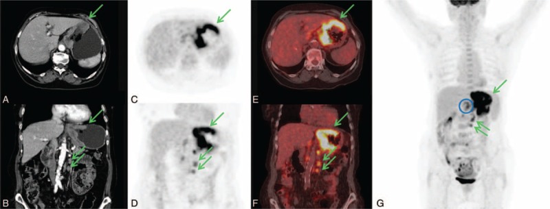
FIGURE 1.

An 83-years-old woman with diagnosis of signet ring cell carcinoma obtained by cardias biopsy. CECT axial and coronal images (A, D) showed regular and diffuse thickening larger than 10 cm in the upper part of the stomach and in left paraortic lymphnodes (green arrows). 18F-FDG PET/CT axial and coronal PET and fused images (B, C, E, F) showed the gastric lesion (SUVmax 13.3) and the left paraortic lymphnodes (SUVmax 7.2) (green arrows). Furthermore, 18F-FDG PET/CT detected celiac lymphnodes involvement (SUVmax 5.1) as is better showed in MIP image (blue circle). CECT = contrast enhancement computed tomography, 18F-FDG PET/CT = fluorine-18 fluoro-2-deoxy-d-glucose positron emission tomography/computed tomography, SUV = standardized uptake value.
