
FIGURE 2.
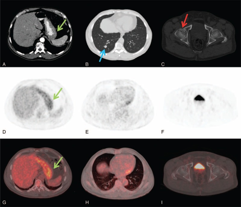
A 54-years-old man with intestinal type gastric carcinoma. CECT axial images showed localized gastric wall thickening (A, green arrow), a right lung nodule of 15 mm suspected for metastases (B, blue arrow) and a osteolytic lesion in the right femoral head doubtful for herniation pit (C, red arrow). 18F-FDG PET/CT axial PET and fused images confirmed the gastric lesion with SUVmax of 4.8 (D, G) but did not showed any 18F-FDG uptake in the lung nodule (E, H) and in the right femoral head (F, I). CECT = contrast enhancement computed tomography, 18F-FDG PET/CT = fluorine-18 fluoro-2-deoxy-d-glucose positron emission tomography/computed tomography, SUV = standardized uptake value.