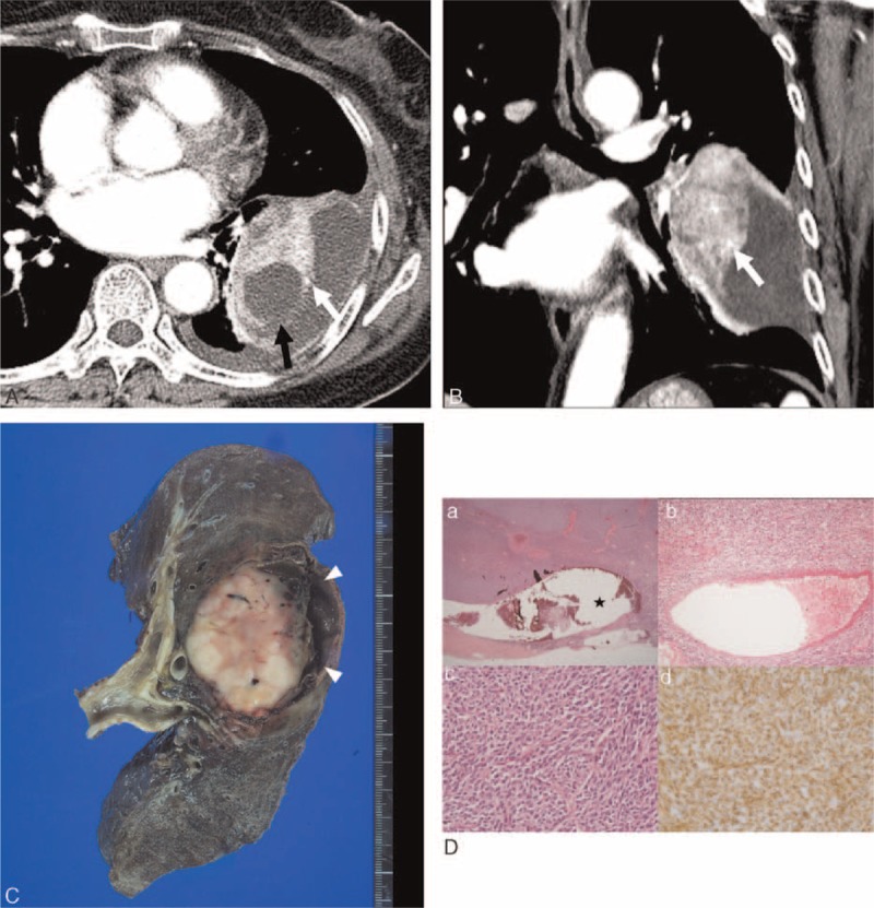
FIGURE 3.

Computed tomography (CT) images obtained in a 58-year-old woman with monophasic primary pulmonary synovial sarcoma. (A and B) Conventional transverse mediastinal CT image (5-mm thick) obtained at the level of the left pulmonary vein, and coronal mediastinal CT image (5-mm thick) obtained at the level of the left upper pulmonary vein. CT images show a 7.7-cm-sized heterogenously enhancing mass in the left lower lobe with circumscribed margin and internal cystic or necrotic portion, and an intratumoral vessel (white arrows in A and B). Note the obliterated tumor margin (black arrow in A) and pleural effusion, indicating rupture of the tumor. FDG PET reveals an FDG-avid lung mass with a maxSUV of 3.8 (not shown). (C) Pneumonectomy was performed and the photograph shows the creamy white and soft solid portion with focal hemorrhage. The cystic portion is filled with a blood clot (arrowheads). (D) On histologic analysis, (a) the periphery of the tumor shows cystic change (★); (b) a thick-walled blood vessel is present in the center of the tumor; (c) the tumor has elevated cellularity and is composed of short spindle-shaped cells with hyperchromatic nuclei, and high mitotic activity (hematoxylin–eosin stain, original magnifications 12.5, ×100, ×400, respectively); and (d) the tumor cells are positive for vimentin by immunohistochemistry. FDG PET = 18F-fluorodeoxyglucose positron emission tomography, maxSUV = maximum standardized uptake value.
