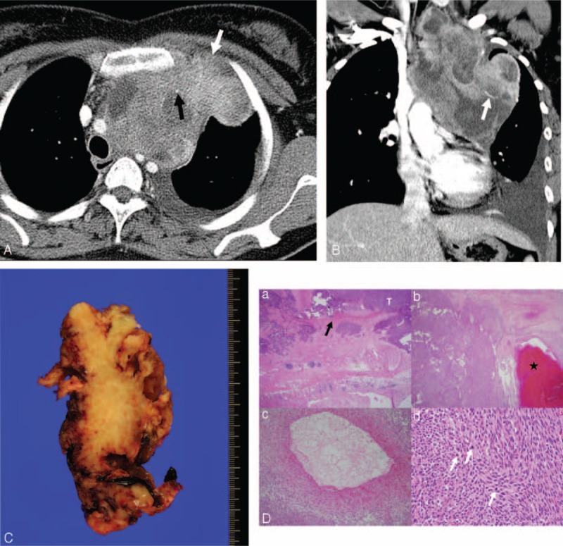
FIGURE 5.

Computed tomography (CT) images obtained in a 33-year-old woman with monophasic primary pulmonary synovial sarcoma in the mediastinum. (A and B) Conventional transverse mediastinal CT image (5-mm thick) obtained at the level of the upper trachea, and coronal mediastinal CT image (5-mm thick) obtained at the level of the superior vena cava. CT images show a 12.8-cm heterogenously enhancing mass with lobulated margin and an internal cystic or necrotic portion that extends to the thoracic inlet. Chest wall invasion (white arrow in A) and pleural effusion are evident. Note the punctate calcification (black arrow in A) and intratumoral vessel (white arrow in B). FDG PET shows an FDG-avid mediastinal mass with a maxSUV of 4.9 (not shown). (C) Incomplete wide resection was performed because of severe adhesion with the left innominate vein and chest wall. The photograph shows the cut surface of the mass with a multifocal hemorrhage. (D) On histopathologic analysis, (a) the tumor (T) invades the pleural wall (black arrow); (b) it shows cystic change with hemorrhage (★); (c) a 1-mm-diameter blood vessel can be seen in the tumor; and (d) the tumor contains hypercellular short spindle-shaped cells with high mitotic activity (white arrows) (hematoxylin–eosin stain, original magnification ×12.5, ×12.5, ×100, ×400, respectively). FDG PET = 18F-fluorodeoxyglucose positron emission tomography, maxSUV = maximum standardized uptake value.
