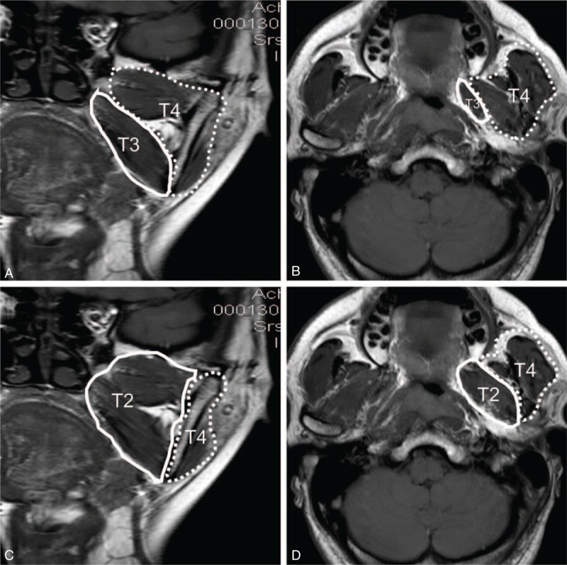
FIGURE 1.

Coronal (A and C) and axial (B and D) images of T1-weighted fast spin echo imaging. According to guidelines for NPC from the seventh edition of the American Joint Committee on Cancer staging system (C and D), the anatomical masticator space includes the medial space (medial and lateral pterygoid muscle, solid line) and the lateral space (temporalis and masseter muscle, dotted line), and tumors involving the medial or lateral part are classified as T4. In contrast, the 2008 Chinese NPC staging system subdivides the masticator space into 2 independent spaces (A and B), including the medial part (only medial pterygoid muscle, solid line) and the lateral part (both lateral pterygoid muscle and other masticatory muscles, dotted line), and tumors involving the medial part are staged as T3 while those involving the lateral part are staged as T4. NPC = nasopharyngeal carcinoma.
