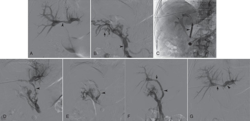
FIGURE 2.

(A) Direct portogram showed normal branches of PV (arrowhead); the main PV was not displayed. (B) A catheter was introduced to traverse the occlusion segment, and entered the superior mesenteric vein (arrowhead); venogram showed rich collateral circulations (arrow), but the main PV was not displayed. (C) A balloon catheter (arrowhead) was used to dilate occluded main PV. (D) Portogram following balloon angioplasty showed multiple filling defect (arrowhead) in the main and right branch of PV, as well as rich collateral circulations. (E) Portogram following stent placement and transcatheter embolization showed multiple filling defect in the stent and little hepatopetal blood flow (arrowhead). (F) Portogram following 24 hours of percutaneous thrombolysis showed normal hepatopetal blood flow (arrowhead) in stent without filling defect; the proximal stent located in the right branch of PV (arrow); the right branch was clearly displayed, while the left branch was not displayed. (G) Intrahepatic portal venogram showed patency of the left branches (arrowhead) and right branches (arrow). PV = portal vein.
