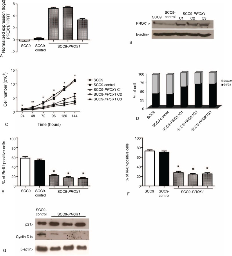
FIGURE 2.

PROX1 overexpression inhibits cell proliferation. PROX1 mRNA expression levels by qRT-PCR in SCC9 cells, SCC9-control, and 3 constitutively PROX1-expressing cell clones (A). Representative Western blot analysis in the cell lines previously described. The β-actin was shown as an internal control (B). PROX1 overexpression was observed in the mRNA and protein levels. Proliferation curves (C), cell cycle analysis by flow cytometry (D), assay measuring BrdU (E), and Ki-67 expression (F) demonstrate that PROX1-overexpressing cells have statistically decreased proliferation compared with control cells (P < 0.001). Western blot for expression of p21 and cyclin D1 (G). PROX1-overexpressing cells showed a decrease in cyclin D1 expression compared with SCC9 and SCC9-control. BrdU = Bromodeoxyuridine, PROX1 = prospero homeobox 1.
