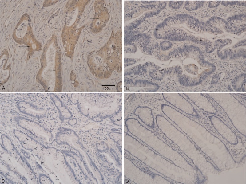
FIGURE 1.

Immunohistochemical staining of XRCC2 expression in CRC and adjacent normal mucosal tissues (×200 magnification). There was (A) positive and (B) weakly positive XRCC2 expression in primary CRC. There was negative XRCC2 expression in (C) CRC and (D) normal colorectal mucosa tissue. Arrows indicate XRCC2-positive cells. The scale bar represents 100 μm.
