
Fig. 4. A 63-year-old male with TIA.
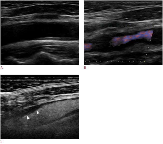
A. The gray-scale sonogram of the origin of the internal carotid artery fails to detect a completely echolucent plaque. B. Directional-eFLOW imaging raises suspicion of filling defects in both sides of the vascular lumen. C. The respective contrast-enhanced ultrasonography image clearly depicts the complete filling of the lumen with microbubbles except for a part of the proximal wall, which is occupied by a homogeneously echolucent plaque (arrowheads). TIA, transient ischemic attack.