
Fig. 5. A 68-year-old female with near-occlusion stenosis of the internal carotid artery (ICA).
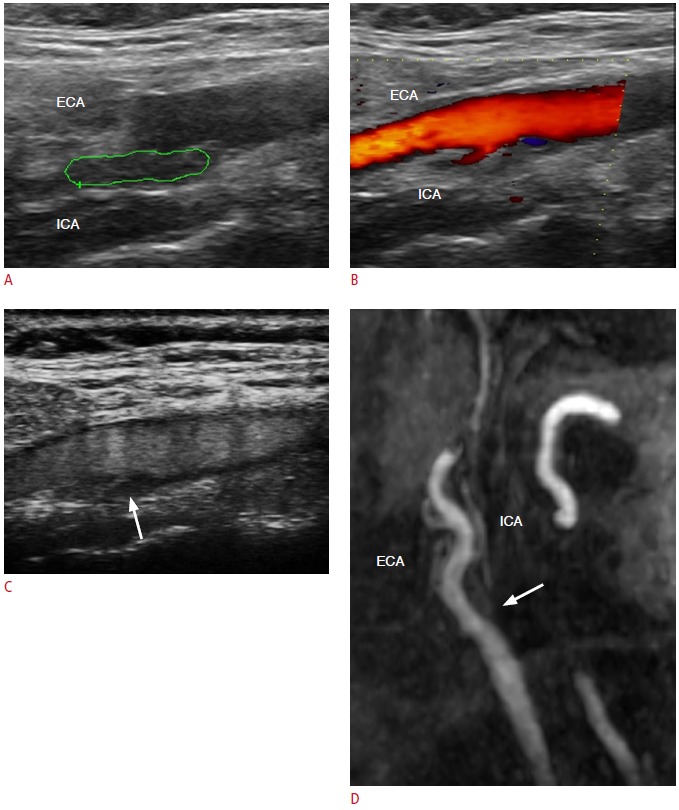
A. The gray-scale sonogram demonstrates an ICA, which is filled with a hypoechoic material (green line represents an ROI for an echogenicity histogram). ECA, external carotid artery. B. The color Doppler technique reveals limited flow signals in the origin of the vessel, which are absent more distally, showing evidence of occlusion. C. Contrast-enhanced ultrasonography demonstrates normal contrast uptake of the common and external carotid artery, while there are moving microbubbles inside the residual lumen towards the periphery of the ICA, posing the diagnosis of its near-occlusion stenosis (arrow). D. Contrast-enhanced magnetic resonance angiography proves the presence of a contrast agent inside the internal carotid, confirming the diagnosis of near-occlusion stenosis (arrow). ROI, region of interest.