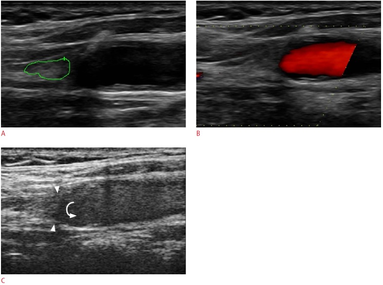
Fig. 6. An 82-year-old male with the occlusion of the right internal carotid artery (ICA).

A. The gray-scale sonogram shows the echogenic material filling the lumen of the ICA (green line represents an ROI for an echogenicity histogram). B. The color Doppler technique reveals no color flow signals inside the internal carotid showing evidence of obstruction. C. Contrast-enhanced ultrasonography confirms the diagnosis with greater confidence as there are no microbubbles detected inside the obstructed vessel (arrowheads). The microbubbles could be seen moving in a revolving pattern at the site of obstruction (curved arrow). ROI, region of interest.
