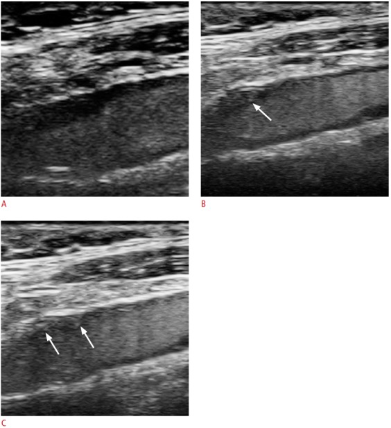
Fig. 8. A 63-year-old male with TIA and a carotid plaque with intraplaque neovascularisation.

A-C. Images of this patient are also presented in Fig. 4. This figure presents a series of images with contrast-enhanced ultrasonography, which were captured at time intervals of several seconds. Of note are the contrast microbubbles, which are seen entering the plaque and represent intraplaque neovascularisation (arrows). TIA, transient ischemic attack.
