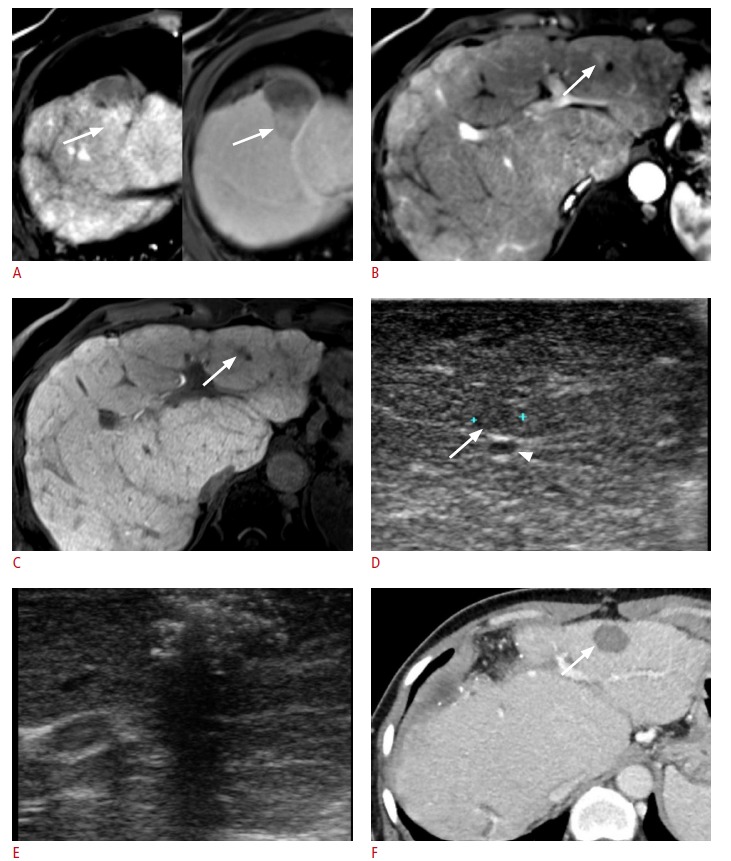
Fig. 2. A 63-year-old female with Budd-Chiari syndrome and hepatocellular carcinoma (HCC) previously treated with transarterial embolization.

A. The arterial (left) and portal (right) phases of contrast-enhanced dynamic magnetic resonance images show the local tumor progression of HCC (arrows) in segment IV of the liver. Surgical resection was planned to treat this tumor. B, C. Preoperative magnetic resonance images show another tiny lesion (arrows) highly suspicious for HCC in segment III of the liver as a hypervascular nodule on arterial-phase imaging (B), and as hypointensity on hepatobiliary-phase imaging (C). D. Intraoperative ultrasonography (IOUS) shows the lesion in segment III near the left hepatic vein (arrowhead) as an isoechoic nodule (arrow). E. Intraoperative radiofrequency ablation was performed for the hepatic nodule with IOUS guidance. F. Follow-up computed tomography shows complete tumor ablation (arrow).
