
Fig. 3. A 70-year-old male with hepatocellular carcinoma (HCC).
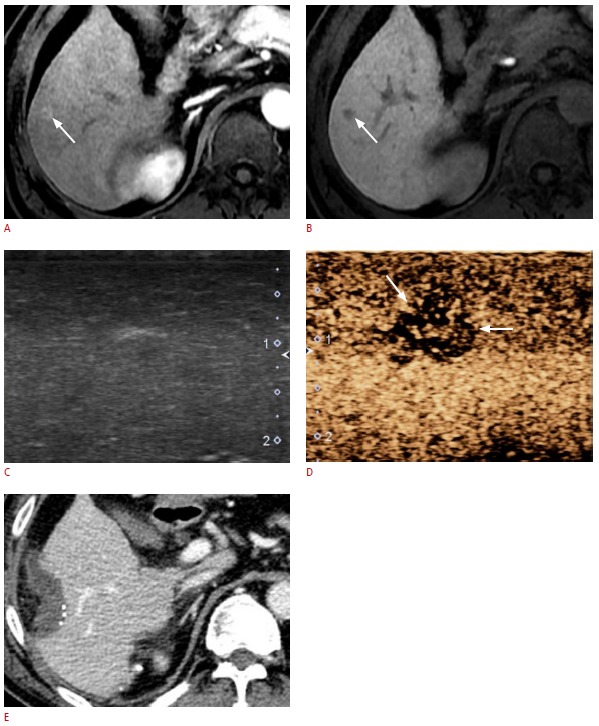
A, B. A preoperative magnetic resonance image shows a 0.8-cm nodule (arrows) in segment VI of the liver as a hyperenhancement on the hepatic arterial phase (A), and as hypointensity on the hepatobiliary phase (B). C, D. Gray-scale intraoperative ultrasonography (IOUS) (C) was not able to find the non-palpable hepatic nodule detected on preoperative magnetic resonance imaging. However, in the Kupffer-phase images of contrast-enhanced IOUS using Sonazoid (D, same scanning plane as C), the nodule (arrows) is clearly distinguished from the background parenchyma as an area of hypoechogenicity. This lesion was pathologically confirmed as HCC. E. Immediate follow-up computed tomography shows that the tumorectomy was successful.