
Fig. 3. A 35-year-old female with lower abdominal discomfort.
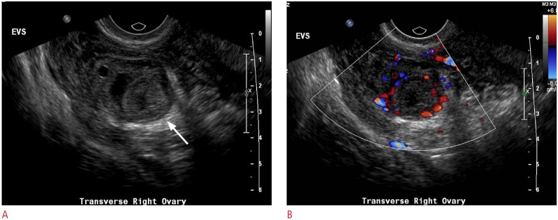
Gray-scale (A) and color (B) sonograms of the right ovary show a round, thick-walled structure (arrow) with a peripheral ring of vascularity on color imaging, consistent with a corpus luteum.