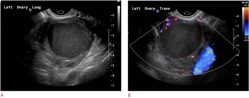
Fig. 7. A 24-year-old female with left pelvic pain.

Gray-scale (A) and color (B) sonograms from a woman with known endometriosis showing peripheral vascularity around a cystic mass with nearly homogenous low-level internal echoes: a typical appearance of an endometrioma.
