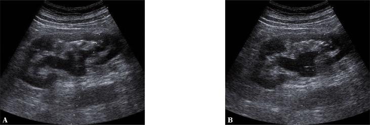
Fig. 9.
A, B. Dilatation of the pelvicalyceal system. The dilatation of the renal pelvis is not accompanied by the widening of the ureter – ultrasound presentation of the dilatation of the ureteropelvic junction. B. A rounded tip of a nephrostomy catheter is visible in the region of the renal pelvis

