Figure 1.
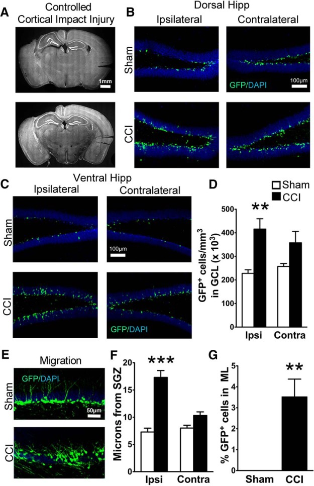
Transgenic POMC-EGFP mice demonstrate CCI-induced neurogenesis and increased dispersion of immature granule cells. A, Representative images of the extent of cortical damage 2 weeks following CCI. The noninjured (contralateral) sides are marked by notches in the cortical tissue placed during processing. B, C, Representative images of GFP+ cells in the dorsal (B) and ventral (C) hippocampus of sham and CCI-treated mice 2 weeks after surgery, both ipsilateral and contralateral to injury. D, CCI-treated mice had more GFP+ cells on the injured hemisphere compared to sham mice (**p = 0.00013, n = 7 and 8 mice/group). E, Representative images of GFP+ cell dispersion in the granule cell layer of the ipsilateral dentate gyrus. F, CCI-treated mice had increased cell migration away from the SGZ on the injured hemisphere compared with sham mice (***p = 0.00018, n = 6 mice/group; white and black bars represent sham and CCI-treated mice, respectively). G, A greater percentage of GFP+ cells from CCI-treated mice migrated into the molecular layer (ML) of the ipsilateral dentate gyrus (**p = 0.002811, n = 6 mice/group).

