Figure 1.

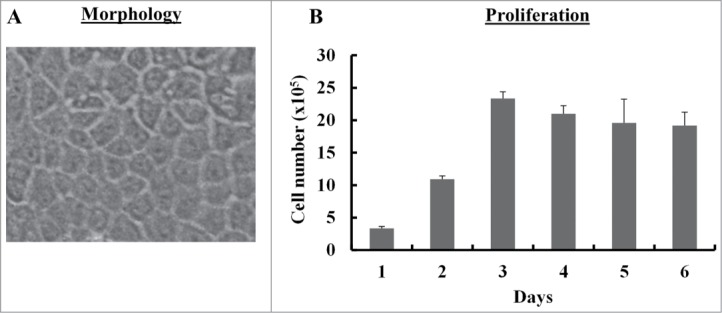
Morphology and proliferation of SUCECs: SUCECs were isolated from the umbilical cords of near term pigs (n = 3) by collagenase treatment. (A) SUCECs displayed characteristic epithelial cell like cobblestone morphology. A representative epithelial colony observed 7–8 d after initial plating of umbilical cord cells is shown. (B) SUCECs (2 × 10⁵/well) were plated in a 6-well plate and their proliferation was measured over a 6 day period by counting the viable cells by trypan blue dye. Data are expressed as mean from triplicates ± SD.
