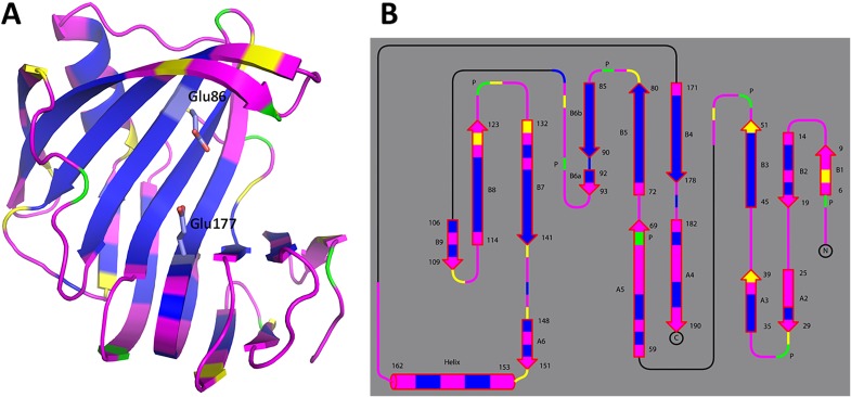
Fig. S4.
Backbone H/D exchange patterns for pD6.2 depicted with the enzyme in diagram representation (A) and as topology (B). Magenta corresponds to fully exchanged main-chain amides (D atom occupancy of 0.60–1.00), yellow is for partially exchanged (D occupancy of 0.00–0.60), and blue indicates nonexchanged NH (D occupancy of −0.56 to 0.00). Proline residues lack the main-chain amide's hydrogen and are depicted in green.

