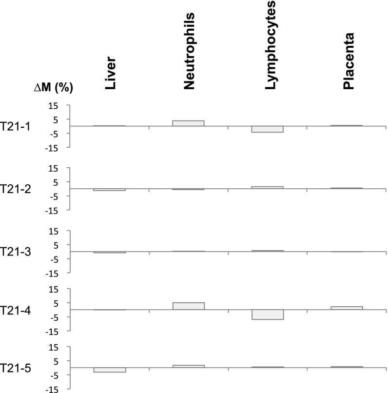
Fig. S1.
∆M values across different tissues for pregnant women each carrying a fetus with trisomy 21 (T21). The methylation markers on all of the autosomes except chromosome 21 were randomly divided into two sets, namely, set A and set B. The randomization was implemented using a series of random numbers (ranged from 0 to 1) generated by a computer. A marker associated with a random number less than 0.5 was assigned to set A; otherwise, it would be assigned to set B. In this analysis, set A included markers originating from chromosomes 1, 2, 4, 5, 6, 8, 12, 14, 15, 17, and 22, and set B included markers originating from chromosomes 3, 7, 9, 10, 11, 13, 16, 18, 19, and 20. Plasma DNA tissue mapping was conducted using each set of markers. The ∆M values shown represent the difference in contributions of a particular tissue to plasma DNA using markers in sets A and B.

