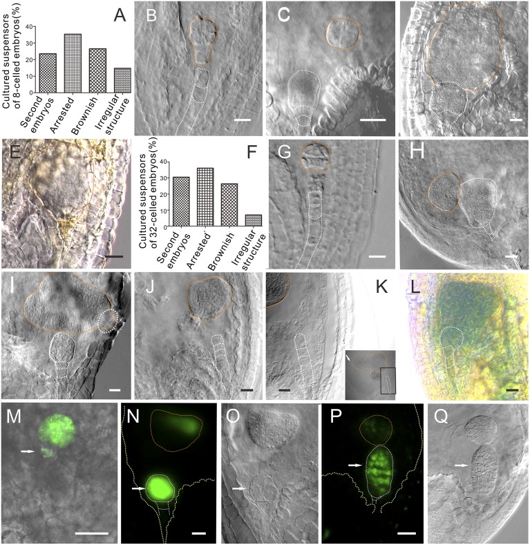
Fig. 1.
The suspensor and embryo proper development after in vivo laser ablation. (A) The statistics of the suspensor development after laser ablation of an eight-celled embryo. (B) The eight-celled embryo after ablation, showing the embryo with two suspensor cells. (C) The laser-ablated eight-celled embryo after a 3-d culture. Note that the suspensor cells were completely removed from the embryo. The top suspensor cell has developed into an embryo (n = 14). (D) The laser-ablated eight-celled embryo developed into a big embryo without further differentiation after a 5-d culture (n = 39). The top suspensor cell has developed into a globular embryo with normal structure and morphology. (E) The brownish suspensor cannot be observed clearly. (F) The statistics of the suspensor development after laser ablation of a 32-celled embryo. (G) The laser-ablated 32-celled embryo after a 3-d culture. The suspensor cells were completely removed. Note the bottom morphology of the embryo. (H) The laser-ablated 32-celled embryo after a 3-d culture (n = 74). (I) The laser-ablated 32-celled embryo after a 5-d culture. The embryo was already differentiated with a clear apical–basal axis. The white dotted-line circle indicates the remnant cells of a suspensor (n = 47). (J) The heart-stage embryo after ablation. (K) The laser-ablated heart-stage embryo after a 3-d culture. The image is an enlargement of the part in the black box. Arrow shows the orientation of embryo with reference to the suspensor (n = 66). (L) The embryo-proper marker ABI3 expressed in the embryo derived from the suspensor cell. (Scale bars, 20 μm.) (M–O) DRN::GFP expressed in both embryos. (O) The differential interference contrast (DIC) image of the same embryos in N. Note that the GFP signal appeared only at the upper part of the heart-shaped embryo. (P) ATML1::H2B:GFP expressed in both embryos. (Q) The DIC image of the same embryos in P. Arrows indicate suspensor-derived embryos in vivo. (Scale bars, 20 μm.)

