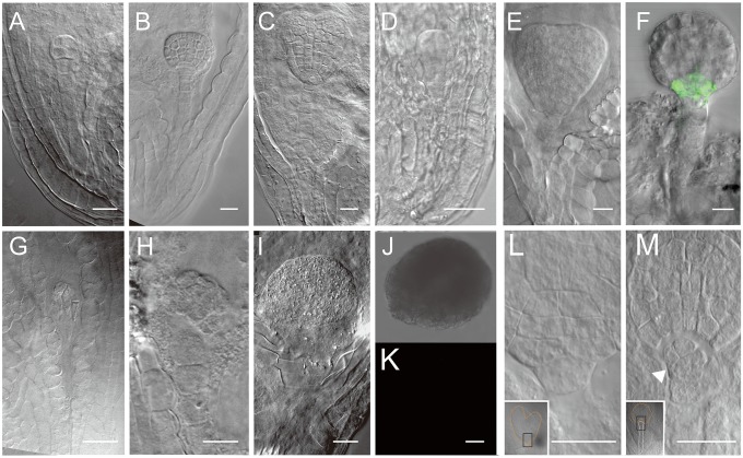
Fig. 2.
Hypophysis formation after laser ablation of suspensor and its role in embryo development. (A–C) Normal embryogenesis as control, showing hypophysis formation and typical morphology of embryo basal part. (D and E) Embryos with remaining suspensor cells, showing that the hypophysis formation and morphology of the embryo basal part is as normal as control. (F) The expression of WOX5 in globular embryo with suspensor cells. (G–I) Embryo without suspensor cells attached, showing the absence of the hypophysis and unique morphology of the embryo basal part. (J and K) The WOX5 do not express in the embryo without suspensor cells attached. (L and M) The magnification of the black boxes. (L) The hypophysis developed normally in the embryo with remnant suspensor cells (n = 26); note the typical cell organization there (dotted line). (M) The embryo proper without suspensor remnants lacked a hypophysis after 3-d culture. Note the first suspensor cell that started to divide (arrowhead) (n = 56).

