Fig. 4.
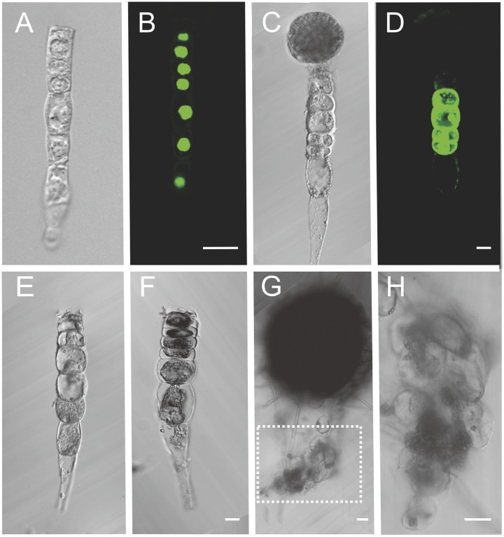
The in vitro culture of isolated suspensors from Arabidopsis and Brassica embryos. (A) The Arabidopsis suspensor of the ZC1::NLS: GFP transgenic lines after laser ablation. (B) The GFP signal of the suspensor in A. (C) The Brassica zygotic embryo after ablation. (D) The FDA (fluorescein diacetate) staining shows the suspensor cell viability. (E) The Brassica suspensor of zygotic embryo after ablation. (F) The suspensor cells of E show cytoplasm shrunken after a 7-d culture (n = 87). (G) The embryo and suspensor development after breaking the connection between them in the medium with hormone. The suspensor cells develop into a cell cluster, but not an embryo. (H) The enlargement of the cell cluster in the box of G. The cells are loosely organized. (Scale bars, 20 μm.)

