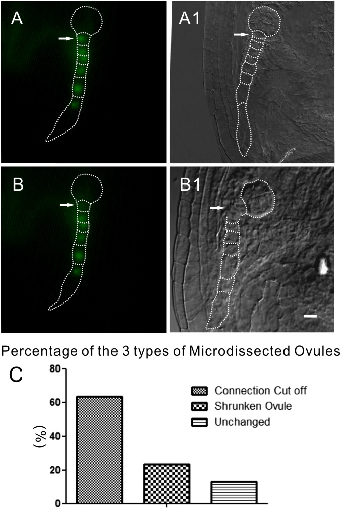
Fig. S2.
In vivo living cell laser microdissection system for removing the connection between suspensors and embryo propers of living embryos. (A and A1) Embryo before laser microdissection. The laser beam usually targets the uppermost suspensor cell (arrows). (B and B1) Embryo after ablation (not the same embryo as in A and A1). The fluorescence in the first cell of the suspensor is eliminated. Arrows indicate the laser-ablated cell. (C) The different percentages of three types of laser-ablated ovule (n = 77). (Scale bars, 20 μm.)

