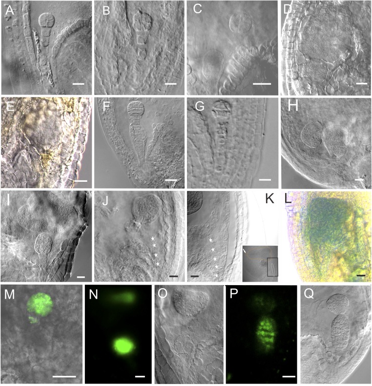
Fig. S5.
The suspensor and embryo proper development after in vivo laser ablation: the images of embryos in Fig. 1 without outlines. (A) The WT eight-celled embryo. (B) The eight-celled embryo after ablation, showing the embryo with two suspensor cells. (C) The laser-ablated eight-celled embryo after a 3-d culture. Note that the suspensor cells were completely removed from the embryo. The top suspensor cell has developed into an embryo (n = 14). (D) The laser-ablated eight-celled embryo developed into a big embryo without further differentiation after a 5-d culture (n = 39). The top suspensor cell has developed into a globular embryo with normal structure and morphology. (E) The brownish suspensor cannot be observed clearly. (F) The WT 32-celled embryo. (G) The laser-ablated 32-celled embryo after a 3-d culture. The suspensor cells were completely removed. Note the bottom morphology of the embryo. (H) The laser-ablated 32-celled embryo after a 3-d culture (n = 74). (I) The laser-ablated 32-celled embryo after a 5-d culture. The embryo was already differentiated with a clear apical–basal axis (n = 47). (J) The heart-stage embryo after ablation. (K) The laser-ablated heart-stage embryo after a 3-d culture. The image is an enlargement of the part in the black box. Arrow shows the orientation of the embryo with reference to the suspensor. Stars indicate suspensor cells (n = 66). (L) The embryo-proper marker ABI3 expressed in the embryo derived from the suspensor cell. (Scale bars, 20 μm.) (M–O) DRN::GFP expressed in both embryos. (O) The DIC image of the same embryos in N. Note that the GFP signal appeared only at the upper part of the heart-shaped embryo. (P) ATML1::H2B:GFP expressed in both embryos. (Q) The DIC image of the same embryos in P. (Scale bars, 20 μm.)

