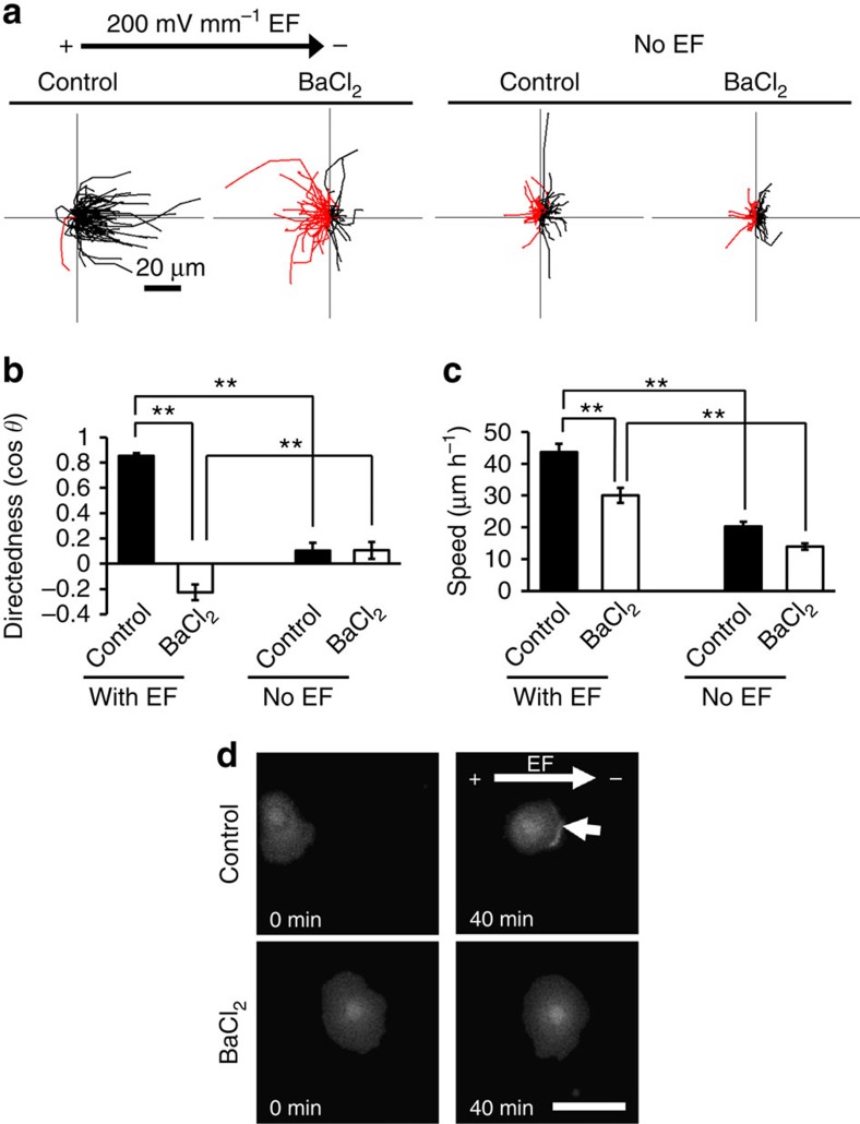
Figure 3. Barium chloride treatment abolished galvanotaxis.
(a) Cells treated with BaCl2 lost galvanotaxis. Black and red lines indicate trajectories of cells migrated toward cathode and anode side, respectively. n=100 cells for each group, confirmed in two other replicates. (b) Directedness values (cos θ) confirm loss of directedness. n=100 cells for each group, confirmed in 2–3 other replicates. (c) BaCl2 treatment significantly inhibited migration speed. n=100 cells for each group, confirmed in 2–3 other replicates. (d) BaCl2 treatment prevented asymmetric accumulation of PIP3 to the leading edge. hTCEpi cells were transfected with pcDNA3-Akt-PH-EGFP plasmid DNA. Fluorescence of Akt-PH-EGFP was recorded by fluorescence microscope. Arrow indicates PIP3 accumulation in cathode-facing side of control cells. Scale bar, 50 μm. BaCl2 was used at 500 μM. EF=200 mV mm−1. Statistical analysis was performed by the Student's t-test. Data represented as mean±s.e.m. **P<0.01.

