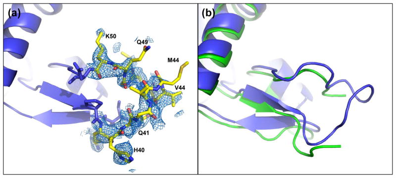
Figure 2.
D-loop conformation. (a) 2FO-FC electron density (0.8 σ) of the D-loop of wide-open state profilin:actin covering residues 41-49. (b) Overlay of the wide-open state profilin:actin (blue) with the partially modeled D-loop of nonexchanging-closed actin (green). The modeled loops are shifted, but show a similar overall structure.

