Figure 2.

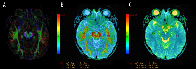
The regions of interest (ROI) positioning in the inferior longitudinal fasciculi (ILF). (A): Color-coded directional map weighted with FA; (B): FA map; (C): ADC map.

The regions of interest (ROI) positioning in the inferior longitudinal fasciculi (ILF). (A): Color-coded directional map weighted with FA; (B): FA map; (C): ADC map.