Figure 2.
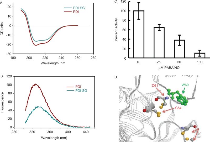
S-glutathionylation of the active-site cysteines on PDI alters protein structure. Spectroscopic analysis of native (red) and PABA/NO+GSH-treated (green) PDI in vitro using circular dichroism (A) and tryptophanyl fluorescence (B) of purified protein. The enzymatic activity of PDI was assessed using the insulin turbidity assay (C). According to the published crystal structure (Tian et al., 2006) and (D), the relative positions of the PDI C61 and 64 and W60 are depicted using Ras Mol 2.7.4.2 (http://rasmol.org last accessed February 18 2011). From Townsend et al., (2009b).

